

Rechercher


Accueil > La Recherche > Axes & Equipes > Bio Nano Imagerie > Equipe : Biophotonique > Thème : Biophotonique
Photonic Biosensors
publié le , mis à jour le
Next generation biosensors will require significant improvements in sensitivity, specificity and parallelism in order to meet the future needs of a variety of fields including in vitro medical diagnostics, pharmaceutical discovery and drug or pathogen detection. The interface between biological molecules and inorganic surfaces is a key issue for the development of devices based on biomolecular recognition. In particular, the efficiency of biosensing devices is directly determined by the specific surface adsorption of targeted biomolecules.
Our objective is developing new sensing substrates presenting high binding selectivity and sensitivity for biological molecules. Miniaturization keeping at the same time large sensing area by assuring high specificity of the sensing surface is the challenge of nowadays research in biosensing. Nanostructured semiconductor substrates are ideal candidates for photonic based molecular detection as they present unique possibilities to support device functionality that includes strongly confined and localized light emission. Miniaturization and large sensing area is assured by nanostructured subtstrates as photonic crystals (PhC) and high specificity can be achieved by their proper functionalization with peptides produced by a biotechnological method called "phage display". We successfully elaborated the peptide sequences for numerous semiconductors (GaN, InP, GaAs, ZnSe, InAs, Si, etc.).2 Specific localization of peptides and reversibility of functionalization was demonstrated and the interactions between the surfaces and its specific peptide have been measured. Peptide –route functionalization was found to preserve enzyme activity and its secondary structure while adsorption on a ZnSe ATR-crystal.3 Use of the peptides sequence found for silicon for sensing and other applications is now under EU patenting. The specific adhesion of these peptides is used for the controlled placement of biomolecules in porous silicon microcavities that are further used for optical biosensing.4
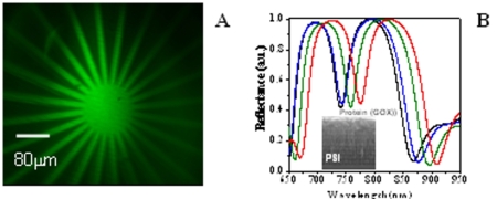
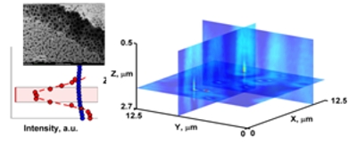
One of the main achievements of our group was the demonstrated fluorescence enhancement via porous silicon mirrors and microcavities.5 This work was highlighted in the article : "Silicon mirrors enhance fluorescence" by Hank Hogan in Biophotonics International, in 2007 and the finding initiated all the research of the team aiming to develop smart microscopic substrates for luminescence enhancement in bioimaging. Moreover, infiltrated protein acts as an internal two-photon-excited fluorescence emitter and second harmonic generator, enabling the in-depth visualization of the porous silicon microcavity by non linear optical microscopy.6
Recently we have obtained encouraging results by observing significant enhancement in the linear and/or non linear optical responses of biomolecules, when embedded in simple 1D photonic structures, compared to unstructured surfaces (PSi vertical micro-cavities and GaAs/AlGaAs structure). Amplification of linear and nonlinear optical responses in photonic crystals makes them ideal candidates for the development of miniaturized biosensors for molecular detection with extreme sensitivity (femtomolar).7 Hence, we plan to continue the development and the study of photonic structures optimized for biosensing applications. Moreover, the observed unique intrinsic luminescent properties of the porous silicon structures can be further exploited as "microscopic biosensors" based on the luminescence response of individual pores when molecules are adsorbed, contrary to the previous sensing methods where detection was based on a macroscopic spectral response of the whole structure.
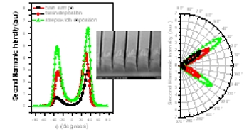
We perform photonic studies to understand the optical behaviour of the bacteriorhodopsin (in purple membranes) that is a natural photonic crystal and a light-driven proton pump in the membrane of the Halobacterium Salinarium. Our optical waveguide lightmode spectroscopy studies provided valuable information’s of the surface charges’ effect on the assemblage of these membranes. Furthermore, non-linear optical measurements at different polarizations revealed an optical chirality of oriented purple membrane films.8
Collaborations : A. M. Malvezzi (Universita di Pavia Italy), S. Lourdoudoss (KTH Sweden), C. Sibilia (Universita di Roma La Sapienza, Italy), E.Perez, G. Palestino (Universidad Autonoma San Luis Potosi, Mexico), V. Agarwal (Universidad Autonoma del Estado de Morelos, Mexico), L. Zimanyi, G. Varo (Biological Research Center, Szeged, Hungary)
Our activities in biophotonics were recognized internationally in the last four years by our participation to the (i) Network of European Excellence PHOREMOST "Nanophotonics to realize molecular scale technologies", (ii) an ECOS-Nord Contract (France-Mexico) : "Exaltation of optical signals of complex proteins polyelectrolytes-immobilized on nanostructured surfaces", (iii) TeraHertz Technology Platform of the Region Languedoc-Roussillon (iv) COST - EU Action MP0702 : Towards functional sub-wavelength photonic structures ; (v) COST- EU TD1002 : « European network on applications of Atomic Force Microscopy to NanoMedicine and Life Sciences » and the European technological platform “Photonics 21”.
Related references :
1) C. Gergely, J. Hemmerlé, P. Schaaf, J.K. Heinrich Horber, J.-C. Voegel and B. Senger (2002) Multi-bead-and-spring model to interpret protein detachment studied by AFM force spectroscopy. Biophys. J. 83 : 706-722.
2) E. Estephan, C. Larroque, N. Bec, P. Martineau, F.J.G. Cuisinier, T. Cloitre and C. Gergely (2009) Selection and mass spectrometry characterization of peptides targeting semiconductor surfaces. Biotechnology & Bioengineering. 104 (6) : 1121-1131.
3) M.-b. Saab, E. Estephan, T. Cloitre, C. Larroque and C. Gergely (2010) Peptide Route Functionalization of ZnSe Crystals Preserves Activity and Structure of Proteins while Adsorption. J. Phys. Chem. C 10.1021/jp104974c
4) G. Palestino, V. Agarwal, R. Aulombard, E. Perez and C. Gergely (2008) Biosensing and protein fluorescence by functionalized porous silicon devices. Langmuir 24 (23), 13765–13771
5) G. Palestino, M.B. de la Mora, J.A. del Rio, C. Gergely, E. Perez (2007) Fluorescence tuning of confined molecules in porous silicon mirrors Appl. Phys. Lett. 91, 12, 121909
6) M. Martin, G. Palestino, V. Agarwal, T. Cloitre, L. Zimányi and C. Gergely (2009) Three dimensional spatial resolution of the nonlinear photoemission from biofunctionalized porous silicon microcavity Appl. Phys. Lett. 94:223313-1-3
7) E. Estephan, D. Bajoni, M.-b. Saab, T. Cloitre, R. Aulombard, C. Larroque, L. C. Andreani, M. Liscidini, A. M. Malvezzi and C. Gergely (2010) Sensing by Means of Nonlinear Optics with Functionalized GaAs/AlGaAs Photonic Crystals. Langmuir DOI : 10.1021/la1000792
8) M. C. Larciprete, A. Belardini, C. Sibilia, M.-b. Saab, G. Váró, and C. Gergely (2010) Optical chirality of bacteriorhodopsin films via second harmonic Maker’s fringes measurements Appl. Phys. Lett. 96, 221108
